Wallander,H., Johansson,L. and Pallon,J.(2002): PIXE analysis to estimate the elemental composition of ectomycorrhizal rhizomorphs grown in contact with different minerals in forest soil. FEMS Microbiology Ecology, 39, 147-156.


Abstract
 The aim of this study was to investigate possible interactions between minerals and ectomycorrhizal (EM) mycelia. Fungal rhizomorphs growing in association with apatite and/or biotite were sampled both from a laboratory experimental system (Rhizopogon sp. and Pinus muricata) and from mesh bags buried in forest soil in the field. The elemental composition of the samples was analyzed with particle-induced X-ray emission (PIXE). Many EM rhizomorphs associated with apatite in laboratory systems and in mesh bags contained larger amounts of Ca (mean ranges between 12 and 31 mg Ca g-1) than similar rhizomorphs connected to acid-washed sand (range 0.3-3.5 mg Ca g-1). Ca originating from apatite was deposited as calcium oxalate crystals on the surface of the rhizomorphs. EM mycelium produced in mesh bags had a capacity to mobilize 0.6 mg P kg-1 year-1 from apatite-amended sand (which is 0.04% of the added apatite). A high concentration of K in some rhizomorphs (up to 11 mg K g-1) suggests that these fungi are good accumulators of K and may have a significant role in transporting K to trees.

Keywords: Calcium oxalate; Ectomycorrhiza; Rhizomorph; Particle-induced X-ray emission; Potassium; Weathering』

要旨
 本研究の目的は、鉱物と外生菌根菌糸間の可能性のある相互作用を調べることである。アパタイト(リン灰石)および/または黒雲母と結びついて成長する根状菌糸束が、実験室実験系から(Rhizopogon sp.および Pinus muricata)と野外の森林土壌に埋めた網袋から採取された。試料の元素組成は粒子励起X線分析法(PIXE)で分析された。実験室系および網袋でのアパタイトに伴った多くの外生菌根根状菌糸束は、酸で洗浄した砂に結びついた類似の根状菌糸束(範囲は0.3〜3.5 mg Ca/g)よりも多量のCa(平均範囲は12〜31 mg Ca/g)を含んでいた。アパタイトに由来するCaは、根状菌糸束の表面にシュウ酸カルシウムの結晶として沈殿していた。網袋に生成した外生菌糸は、アパタイトで改良した砂(アパタイトが0.04%添加されたもの)から 0.6 mg P/kg/年を移動させる能力をもっていた。いくつかの根状菌糸束に高濃度のK(11 mg K/gまで)があるのは、これらの菌類がKをうまく濃集する働きをし、樹木にKを輸送するのに重要な役割を持っている可能性を示している。』

1. Introduction
2. Materials and methods
 2.1. Laboratory experiment
 2.2. Field experiments
 2.3. PIXE analysis
 2.4. Scanning electron microscopy (SEM) analysis
 2.5. Statistical analysis
3. Results
 3.1. Laboratory experiments
 3.2. Field-collected samples
4. Discussion
Acknowledgements
References

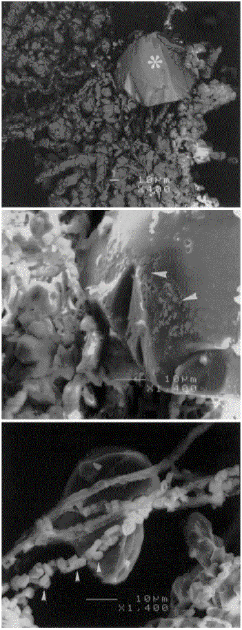
Fig. 3. SEM micrograph of rhizomorphs collected from inside a container with apatite and biotite in a laboratory experiment. Top: an overview of the rhizomorph, which is covered by Ca oxalate crystals and an embedded apatite particle (*), is seen to the right. Middle: close up view of the apatite particle which shows signs of erosion of the surface (arrowheads). Bottom: close up view of fungal hyphae covered by calcium oxalate crystals (arrowheads).

図3.実験室での実験でアパタイトと黒雲母を含む容器内から採取された根状菌糸束のSEM写真。:根状菌糸束の概観で、Caシュウ酸塩結晶に覆われており、また右側に取り囲まれたアパタイト粒子(*)が見える。:表面に浸食の兆しを示すアパタイト粒子の拡大像(矢じり印)。:シュウ酸カルシウム結晶に覆われた菌糸の拡大像(矢じり印)。

〔『Wallander,H., Johansson,L. and Pallon,J.(2002): PIXE analysis to estimate the elemental composition of ectomycorrhizal rhizomorphs grown in contact with different minerals in forest soil. FEMS Microbiology Ecology, 39, 147-156.』から〕


戻る